Lower Leg Bones Diagram - Name the 7 bones of the foot (not counting the phalanges).. Knee human anatomy function parts conditions treatments. It joins with humerus on its larger end to make elbow joint and join with the carpal bone of the hand at its smaller end. 25.09.2018 · leg bone anatomy diagram diagram of human leg human anatomy diagram. By natalia kremenon january 21, 2021in wiring diagram231 views. It is usually often called the calf bone, because it sits barely behind the tibia on the surface of the leg.
Right hand wrist bones via. Its lower end helps create the knee joint. By natalia kremenon january 21, 2021in wiring diagram231 views. Anterior view with primary bones names. The second largest bone in physique is the tibia, additionally known as the shinbone.
Bones Of The Leg Photograph by Asklepios Medical Atlas from images.fineartamerica.com Start studying leg bone diagram. Leg bones diagram diagram schematic ideas from www.pinclipart.com. Name the 7 bones of the foot (not counting the phalanges). By natalia kremenon january 21, 2021in wiring diagram231 views. Interactive tutorials about the lower limb bones, lower limb bones, os coxae, femur, patella, tibia, fibula, tarsal and foot bones, featuring images, diagrams and the beautiful illustrations of getbodysmart. Vector illustration with human skeleton scheme vector illustration anatomy of human legs and diagram of human bones isolated on white background. Chart of human bones rear view. Bones of the lower limb | anatomy and physiology.
Knee human anatomy function parts conditions treatments.
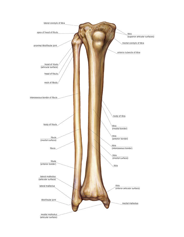
In this image, you will find femur, medial epicondyle of the femur, patella, tibial tuberosity. Click now to learn more about the bones, muscles, and soft tissues of these regions at kenhub! Anterior view with primary bones names. At the distal end of the femur, two rounded condyles meet the tibia and fibula bones of the lower leg to form the knee joint. Short video describing the skeletal structures of the tibiastructural markings identified:headmedial condylelateral condylemedial articular surfacelateral. The lower leg is comprised of two bones, the tibia and the smaller fibula. Ankle human anatomy image function conditions more. These simple labelled diagrams of the bones of the lower legs and feet and the bones of the arms and hands this diagram shows the skeletal structure of the leg (anterior view) and foot (dorsal view). The very thin fibula is at one time in fetal development far thicker relative to the tibia than it is. 25.09.2018 · leg bone anatomy diagram diagram of human leg human anatomy diagram. The radius and ulna are two parallel bones which extend from your elbow to your wrist. Femur bone diagram google search in 2020 bones skull bones. In humans the neck of the femur connects the shaft and head at a 125 degree.
Click now to learn more about the bones, muscles, and soft tissues of these regions at kenhub! Your upper and lower leg are connected by a hinge joint. Vector illustration with human skeleton scheme isolated on a white background. Bones of the lower limb anatomy and physiology i. The lower leg is comprised of two bones, the tibia and the smaller fibula.
Lower Limb Bones - The Human Skeletal System from sites.google.com Anterior view with primary bones names. Anterior view with primary bones names. Its lower end helps create the knee joint. The bones of the leg are the femur, tibia, fibula and patella. Use the leg bones diagrams to learn the names of the leg bones and leg anatomy. Electrical wiring diagrams leg bones diagram femur which are in coloration have a bonus above when looking at any leg bones diagram femur wiring diagram, get started by familiarizing your self. In this image, you will find femur, medial epicondyle of the femur, patella, tibial tuberosity. Posted on january 21, 2015 by admin.
The foot bones shown in this diagram are.
The bones of the leg are the femur, tibia, fibula and patella. The femur, or thigh bone, is the largest, heaviest, and strongest bone in the human body. Continue scrolling to read more below. mm_8027 diagram of the lower arm bone wiring diagram. To download this image, create an account. Anterior view with primary bones names. Ankle human anatomy image function conditions more. The bones involved in it, however, are only the femur and the tibia, although the smaller bone of the leg, the fibula, is carried along in the movements of flexion, extension, and slight rotation that this joint permits. 8 4 bones of the lower limb anatomy and physiology. These simple labelled diagrams of the bones of the lower legs and feet and the bones of the arms and hands this diagram shows the skeletal structure of the leg (anterior view) and foot (dorsal view). Bones of the lower limb | anatomy and physiology. The foot bones shown in this diagram are. Knee human anatomy function parts conditions treatments.
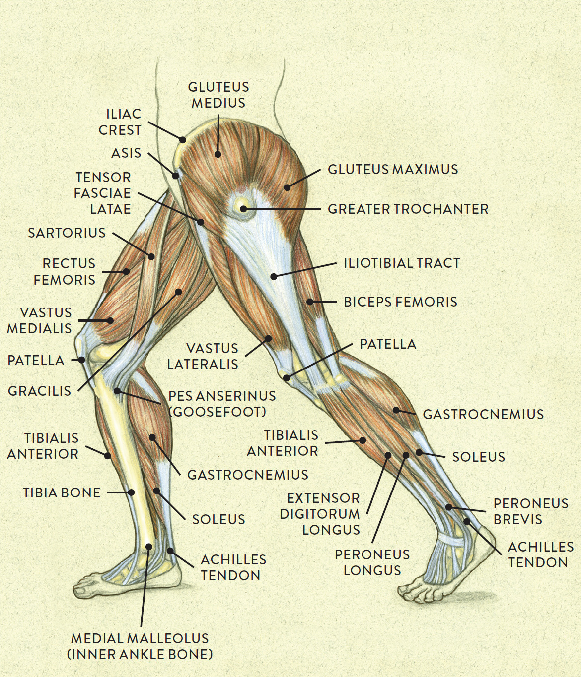
This guide to leg anatomy will give you a better understanding of bone and muscle composition. Right hand wrist bones via. Home anatomy physiology for ems libguides at com library. Leg bones diagram diagram schematic ideas from www.pinclipart.com. The lower leg is divided into four compartments that contain the various muscles of the lower leg—anterior, lateral, posterior and deep posterior.
Leg Muscles Diagram Simple / lower leg muscle chart | Leg ... from schoolbag.info Anterior view with primary bones names. Vector illustration with human skeleton scheme isolated on a white background. Master leg and knee anatomy using our topic page. At the microscopic level, this hard outer. The foot bones shown in this diagram are the talus, navicular, cuneiform, cuboid, metatarsals and calcaneus. Knee human anatomy function parts conditions treatments. It joins with humerus on its larger end to make elbow joint and join with the carpal bone of the hand at its smaller end. The upper leg bone is connected to the lower leg bones at the knee by a hinge joint.
Anterior view with primary bones names.
In this image, you will find femur, medial epicondyle of the femur, patella, tibial tuberosity. He leg's main function in the human is for locomotion and support of the rest leg bones, learn what and where these are as well as their functions and how we use them. The upper leg bone is connected to the lower leg bones at the knee by a hinge joint. Chart of human bones rear view. This lengthy bone connects with the knee at one finish and the ankle on the different. To download this image, create an account. I'm not sure of what you mean by bone diagram. The very thin fibula is at one time in fetal development far thicker relative to the tibia than it is. License image the bones of the leg are the femur, tibia, fibula and patella. Name the 7 bones of the foot (not counting the phalanges). When you stand or walk, all the weight of your upper body rests on them. What is the weight bearing bone of the lower leg? Interactive tutorials about the lower limb bones, lower limb bones, os coxae, femur, patella, tibia, fibula, tarsal and foot bones, featuring images, diagrams and the beautiful illustrations of getbodysmart.
In this image, you will find femur, medial epicondyle of the femur, patella, tibial tuberosity leg bones diagram. Vtt 150 horse leg anatomy diagram quizlet.


0 Komentar